
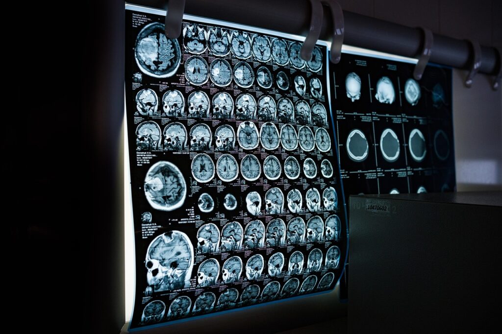
There is a quiet revolution happening inside hospital walls, one that doesn’t involve scalpels or sutures. It is the world of radiology, where beams of invisible energy are turned into maps of the human body. What began as a curious experiment with X-rays over a century ago has grown into the backbone of modern diagnosis, guiding everything from a broken bone to a brain tumor.
The magic lies in the physics. Electromagnetic radiation—X-rays, gamma rays, and their high-energy cousins—passes through flesh and bone, but not equally. Dense tissue like bone absorbs more, while softer organs let more through. That difference, called attenuation, creates the shadows and contrasts that radiologists read like a language. The result is a suite of tools that have become household names: CT scans, MRIs, PET scans, and ultrasound. Each offers a different window into the body, from the sharp cross-sections of a CT to the soft-tissue clarity of an MRI.
But radiology is not just about taking pictures. It has a surgical side, too. Interventional radiologists use imaging to guide tiny instruments through blood vessels, unblocking arteries, cutting off blood supply to tumors, or burning away abnormal tissue with heat. These procedures spare patients the trauma of open surgery, shortening hospital stays and speeding up recovery. A patient who once faced a week of healing now walks out in a day.
The field is also leaping forward with tools that would have seemed like science fiction a decade ago. Functional MRI tracks blood flow to reveal which parts of the brain are working during a thought or a memory. Diffusion tensor imaging maps the brain’s wiring, showing how different regions talk to each other. Spectroscopy goes even deeper, reading the chemical makeup of tissues. And now, artificial intelligence is stepping in as a tireless assistant, scanning thousands of images in minutes to flag suspicious spots that a human eye might miss. The result is faster, sharper, and more accurate diagnoses.
None of this happens in a vacuum. Radiology sits at the intersection of physics, engineering, and medicine, a place where the abstract world of atoms and electromagnetic waves meets the very real needs of patients. It is a field that rewards curiosity and patience, where a single image can change a life. And as the technology keeps evolving, so does our understanding of what it means to be human.
The journey into this realm is not just about machines and algorithms. It is about the people who interpret those images, the patients who place their trust in them, and the discoveries still waiting to be made. Radiology has already reshaped medicine, but the best is yet to come.


![Osun election: Voters turn out early, wait for INEC officials [Video]](https://mediatalkafrica.com/wp-content/uploads/2026/08/Osun-voters-queue-early-for-governorship-election-as-INEC-materials-768x576.jpg)